Saturday, February 28, 2009
Tuesday, February 24, 2009
Music, Memory, and Emotion
Here is my article from ScienceDaily:
Enjoy!
Saturday, February 21, 2009
Tuesday, February 17, 2009
Report on Invisible Images, Attention, Awareness, and Possibilities in Subliminal Messaging
How does an image become invisible?
Bahrami, et. al. used the same method to render images invisible as did a previous team, Fang and He. This method is called flash suppression. It goes as follows: a low-contrast, simple image (of a tool such as a wrench in this study) is shown to one eye while simultaneously, a high-contrast, dynamic noise image is flashed repeatedly to the other eye. In each eye's visual field, the image locations are the same. Subjects of the study were unable to detect the fainter, simpler image, "confirming their complete lack of awareness" that they'd seen it at all - hence, invisibility.
Invisible, but still affecting your brain:
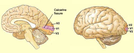
Fang and He found that despite their lack of awareness of the tool images, subjects were nevertheless affected at the neurological level. Using function Magnetic Resonance Imaging (fMRI), they found that the tool images "activated dorsal visual areas along the intraparietal sulcus at almost normal levels, but did not activate ventral visual areas of the lateral occipital complex." This affirms the 'conventional notion' that conscious visual perception is supported by the ventral visual cortex, while 'subconscious' vision (see image below) is supported by the dorsal visual cortex. Essentially, Fang and He found that a subject could 'see' an object with his brain, with almost a completely normal neurological effect on the dorsal side, while remaining completely unaware that he has done so - ventral side unaffected.
Ventral vs. Dorsal pathways illustrated:

Bahrami, et. al. investigate attention’s effect on awareness:
The study noted in the original ScienceDaily article, conducted at Univerity College of London, used the same model as the Fang and He study with low-contrast ‘invisible’ tools and high-contrast noise images. However, they added a focus of attention for their subjects as well. The invisible images and the noise images were both shown in the periphery of the visual field while in the center of the visual field, subjects were asked to observe a stream of letters. They had subjects perform simple tasks in picking certain letters to assure that their attention remained there while taking using fMRI to map the brain’s responses. Bahrami, et. al. had subjects perform a ‘low attention task,’ such as ‘report all Zs’ and a ‘high attention task,’ such as ‘report blue Zs and white Ns.’ See the diagram below for a basic illustration of the study:

Figure 1. A schematic representation of the display used by Bahrami et al. [6].At peripheral locations, one eye views high-contrast, dynamic noise images that are visible to the observer. At some of the same locations, the other eye views low-contrast tool objects that remain invisible. Both eyes view identical letters at the center of the visual field. The complexity of a letter-monitoring task controls the extent to which peripheral images are attended.
The results:
Bahrami, et. al. found that the peripheral ‘invisible’ images still registered in fMRI when subjects were performing low-attention tasks, but not when their attention was drawn away more insistently. In the low-attention task trials, the subjects were able to tell where the noise-images were flashing, and the invisible tool images had a greater affect on the visual cortex. By contrast, the high-attention task rendered the subjects less able to place the location of the peripheral images and showed less fMRI activation from the invisible tool images. So, Bahrami, et. al. found that ‘a neuronal response need not contribute to visual awareness, even though it is enhanced by visual attention. In short, attention does not guarantee awareness.”
Why is this important?
Subliminal messaging (brief flashing images hidden in other media, such as a picture of a Coca-Cola flashed during a projected movie) is illegal in Britain, but not in the USA. There is continued debate as to whether subliminal messaging is an effective advertising tool, or even possibly a tool for controlling the thoughts and opinions of the general public – imagine if subliminal messages could be used to get people to vote for a political candidate, or to encourage the public to approve of an unpopular war. Subliminal messaging has never been proven to ‘work,’ but it remains a fascinating hypothesis. The Fang and He study showed that there is a neurological response to some images even when the viewer remains unaware of having seen them. Bahrami, et. al. showed that attention affects this brain activity. Essentially, if the subject is focusing attentively on one task, even if it is a visually-based task, the brain will register less about an invisible flashed image. This implies that too much attention paid to the ‘overt message’ could conceivably cancel out the subliminal message. Maybe advertisers should only flash images of their products during ‘low-attention’ viewing tasks (such as the boring part of the movie?) instead of the ‘high-attention’ times (extremely complicated chase sequence?). This is simply speculation – these scientific studies cannot be said to solve the question of subliminal advertising once and for all. As Bahrami says, “What our study doesn't address is whether this would then influence you to go out and buy a product. I believe that it's likely that subliminal advertising may affect our decisions -- but that is just speculation at this point."
Bibliography:
F. Fang and S. He, Cortical responses to invisible objects in the human dorsal and ventral pathways, Nat. Neurosci. 8 (2005), pp. 1380–1385.
B. Bahrami, N. Lavie and G. Rees, Attentional load modulates responses of human primary visual cortex to invisible stimuli, Curr. Biol. 17 (2007), pp. 509–513
J. Braun, Vision: Attending the Visible, Curr. Biol. 17 (2007), pp. R202-R203
University College London (2007, March 9). Subliminal Advertising Leaves Its Mark On The Brain. ScienceDaily. Retrieved February 17, 2009, from http://www.sciencedaily.com /releases/2007/03/070308121938.htm
C. Koch and N. Tsuchiya, Attention and consciousness: two distinct brain processes, Trends Cogn. Sci. 11 (2007), pp. 16–22.
Presentation on Grey Matter and First Episode Psychosis
A new study has shown there are grey matter deficits in the brains of children ages 7 - 18 with early onset first episode psychosis (FEP). The study featured patients with schizophrenia, bipolar I disorder, and other psychoses.


Grey Matter
Grey matter is a component of the central nervous system, and contains neural cell bodies. The density of grey matter in a particular area corresponds with various abilities.
The top image shows a T1 MRI differentiating between grey and white matter while the bottom image from the New York Times shows the distinction.
Various diseases and disorders have been traced to a lack or atrophy of grey matter, including schizophrenia, bipolar disorder, dyslexia, and MS. These all show decreased amounts of grey matter in specific areas of the brain. In diseases like schizophrenia atrophy occurs over time and affects various areas of the brain.
Schizophrenia and Bipolar I Disorder
Schizophrenia is a mental disorder characterized by abnormalities in perceptions of reality. There are several characteristics of schizophrenia including delusions, hallucinations, disorganized speech, catatonic behavior, and negative symptoms such as attempted flattening (DSM-IV). In general two of these characteristics, which can not be attributed to any other disease or disorder, must be present for 6 months in order for diagnosis.
Bipolar I disorder is a sub-classification of bipolar disorder. Bipolar disorder is characterized by episodes of either mania, an abnormally elevated mood, or depression. Some sufferers may also have mixed episodes, where both symptoms of mania and depression are present at the same time. Bipolar I disorder is characterized by the presence of a manic episode, whether or not the patient has has a depressive or mixed episode (although these are common in bipolar I patients). This is in contrast to bipolar II sufferers who have only had hypomanic (less severe mania) or depressive episodes. The DSM-IV considers there to be a spectrum of bipolar disorders including, bipolar I and II as well as cyclothomia and bipolar n.o.s. (not otherwise specified).
About this study/Why is it important
While past studies have shown grey matter deficits in older patients and people with later onset FEP this was the first study to look specifically at young patients upon first onset. In addition this study focused on a variety of FEP patients, instead of just schizophrenics, a disease where most of this research is concentrated. The study focused on 121 children and adolescents ages 7 - 18, 70 with early onset FEP and 51 control subjects. The subjects' brains were imaged using MRI technology. Follow up care was provided and the patients were diagnosed. Of these diagnoses 25 patients presented with schizophrenia, 20 with bipolar I disorder, and 25 with other psychoses. While all of these patients showed a decrease in grey matter, patients with schizophrenia and bipolar I disorder showed a specific atrophy in the left medial frontal gyrus. In contrast patients with other psychoses showed smaller bilateral grey matter volumes in the insula and right middle occipital gyrus. The shared pathology between schizophrenia and bipolar I disorder may lead to further investigation into the similarities in the diseases as well as the development of new, more targeted medicines that may prevent future grey matter atrophy.
Bibliography
Jansen, J., Reig, S., Parellada, M., et all. (2008). Regional Grey Matter Volume Deficits in Adolescents with First-Episode Psychosis. Journal of the American Academy of Child and Adolescent Psychology. Vol 47 Issue 11, 1311-1320.
Zipursky, R., Lambe, E., Kapur, S., Mikulis, D. (1998). Cerebral Grey Matter Volume Deficits in First Episode Psychosis. Arch Gen Psychiatry, 55, 540-546.
Gur, R., Turestsky, B., Bilker, W., Gur, R. (1999). Reduced Grey Matter Volume in Schizophrenia. Arch Gen Psychiatry, 56, 905-911.
Harrison, P. (1999). The neuropathology of schizophrenia. Brain, Vol 122, No. 4, 593-624.
Steen, R., Mull C. , McClure, R., Hammer, R., Liebermann, J. (2006). Brain Volume in First Episode Schizophrenia. British Journal of Psychiatry, 188, 510-518.
Monday, February 16, 2009
Saturday, February 14, 2009
Grey Matter and Schizophrenia
Here's the link to the Science Daily article about how lack of grey matter in the brain is linked to schizophrenia and bipolar disorder.
Friday, February 13, 2009
Meditation, Mindfulness and Cognitive Flexibility
Brought to you by Elsevier Journal. Enjoy!
http://www.sciencedirect.com/science?_ob=ArticleURL&_udi=B6WD0-4VH333S-1&_user=2670204&_coverDate=01%2F31%2F2009&_alid=863876619&_rdoc=3&_fmt=high&_orig=search&_cdi=6752&_sort=d&_st=4&_docanchor=&_ct=271&_acct=C000058509&_version=1&_urlVersion=0&_userid=2670204&md5=186be3be4027f8eb20bcd970eaf905b5
http://www.sciencedirect.com/science?_ob=ArticleURL&_udi=B6WD0-4VH333S-1&_user=2670204&_coverDate=01%2F31%2F2009&_alid=863876619&_rdoc=3&_fmt=high&_orig=search&_cdi=6752&_sort=d&_st=4&_docanchor=&_ct=271&_acct=C000058509&_version=1&_urlVersion=0&_userid=2670204&md5=186be3be4027f8eb20bcd970eaf905b5
Tuesday, February 3, 2009

Blindsight Presentation
Blindsight
Definition of Blindsight
The patient TN reported on in this NPR story is the latest in a series of cases of blindsight. This paradoxical and counterintuitive phenomenon refers to the ability of humans with a loss of primary visual cortex to make visual discriminations in their blind visual fields without awareness of the stimuli they are discriminating.

Previous Cases:
This phenomenon was first studied in human patients the 1970s by Oxford University based researchers Lawrence Weiskrantz and Elizabeth Warrington. Their patient GY had extensive damage to his left visual cortex which rendered him functionally blind in his right visual field. They were able to demonstrate GY's capacity to perfectly discriminate the direction of motion within his right visual field. Figure 1 of Weiskrantz's review paper shows the different directions of motion that GY was able to accurately mimic with his arm. The grey area is the impaired hemi-field.
Controversies about the cause of blindsight:
Blindsight is most likely to be due to the use of visual pathways outside of the usual geniculostriate ones, connections that are either subcortical or that go directly to extrastriate areas bypassing primary visual cortex. Some brain researchers have objected that the residual visual function of blindsight could be subserved by fragments or islands of intact striate cortex rather than extrastriate cortex (Weiskrantz, 1995). This is unlikely to be the explanation for GY's motion, wavelength, and emotional expression discrimination capacities because a high-resolution MRI scan reveals only a small patch of striate cortex near the back of the brain on the left side, but he does have some remaining striate cortex.
Why TN is a notable case
The damage to TN's striate cortex is much more extensive than GY's. TN suffered two strokes 36 days apart; the first damaged his occipital cortex unilaterally, and the second destroyed the remaining primary visual cortex in the other hemisphere. Figure 1 of de Gelder et al's paper reporting the case shows the extensive primary visual cortex damage. TN is the only available case in the literature with selective bilateral occipital damage. Yet he can successfully navigate down a long corridor with various barriers set in his way, as demonstrated in the video. His blindsight despite total loss of primary visual cortex effectively refutes the remaining islands of functional visual cortex hypothesis. Extra-striate pathways in humans can sustain sophisticated visuo-spatial skills in the absence of perceptual awareness.
What is blindsight good for?
Blindsight is not demonstrated in every patient with loss of primary visual cortex, but when it is present then it the ability can be cultivated through training for rehabilitation. In the case of TN he was unaware of his residual ability to navigate obstacles using visual information. Behaviorally he was blind across the whole visual field. He walked like a blind man, using his stick to track obstacles and requiring guidance by another person when walking around the laboratory buildings during testing. The researchers were able to demonstrate navigation capacity that he did not know that he still retained in the face of such devastating visual loss. In their quick guide to blindsight for the journal Current Biology Stoerig and Cowey (2007) conclude with the speculation that implicit processes in many domains may always survive when explicit representations are damaged, and therefore that rehabilitation programs could always successfully harness the remaining implicit capacities for restitution.
Bibliography:
de Gelder, B., Tamietto, M., van Boxtel, G., Goebel, R. Sahraie, A., van den Stock, J., Steinen, B.M.C., Weiskrantz, L. & Pegna, A. (2008). Intact navigation skills after bilateral loss of striate cortex. Current Biology, 18, 1128-1129. Link
Lamme, V.A.F. (2006). Zap! Magnetic tricks on conscious and unconscious vision. Trends in Cognitive Science, 10, 193-195.
Rees, G. (1999). Consciousness lost and found. Journal of Psychophysiology, 13, 56-60.
Stoerig, P. & Cowey, A. (2007). Blindsight quick guide. Current Biology, 17, 822-824.
Tamietto, M. & deGelder, B. (2008). Affective blindsight in the intact brain: Neural intrahemispheric summation for unseen facial expressions. Neuropsychologia, 46, 820-828.
Weiskrantz, L. ( 1995). Blindsight - Not an island unto itself. Current Directions in Psychological Science, 4, 146-151. Link to Academic Search Premier
Definition of Blindsight
The patient TN reported on in this NPR story is the latest in a series of cases of blindsight. This paradoxical and counterintuitive phenomenon refers to the ability of humans with a loss of primary visual cortex to make visual discriminations in their blind visual fields without awareness of the stimuli they are discriminating.

Measurement of Blindsight
To get around the lack of visual awareness of blind field stimuli researchers ask their patients to guess whether, where, or which one of a small number of stimuli has been presented within the blind visual field. The types of visual discriminations that have been reported are movement, orientation, wavelength (i.e. , color), spatial localization and combinations of these elementary visual features. Accuracy of responses sometimes reached 90% to 100% in various patients (Weiskrantz, 1995). In addition 'affective blindsight' has been demonstrated: patients can sometimes reliably detect the valence of emotional expressions in the absence of any visual awareness of the faces (Tamietto & deGelder, 2008).
Previous Cases:
This phenomenon was first studied in human patients the 1970s by Oxford University based researchers Lawrence Weiskrantz and Elizabeth Warrington. Their patient GY had extensive damage to his left visual cortex which rendered him functionally blind in his right visual field. They were able to demonstrate GY's capacity to perfectly discriminate the direction of motion within his right visual field. Figure 1 of Weiskrantz's review paper shows the different directions of motion that GY was able to accurately mimic with his arm. The grey area is the impaired hemi-field.
Controversies about the cause of blindsight:
Blindsight is most likely to be due to the use of visual pathways outside of the usual geniculostriate ones, connections that are either subcortical or that go directly to extrastriate areas bypassing primary visual cortex. Some brain researchers have objected that the residual visual function of blindsight could be subserved by fragments or islands of intact striate cortex rather than extrastriate cortex (Weiskrantz, 1995). This is unlikely to be the explanation for GY's motion, wavelength, and emotional expression discrimination capacities because a high-resolution MRI scan reveals only a small patch of striate cortex near the back of the brain on the left side, but he does have some remaining striate cortex.
Why TN is a notable case
The damage to TN's striate cortex is much more extensive than GY's. TN suffered two strokes 36 days apart; the first damaged his occipital cortex unilaterally, and the second destroyed the remaining primary visual cortex in the other hemisphere. Figure 1 of de Gelder et al's paper reporting the case shows the extensive primary visual cortex damage. TN is the only available case in the literature with selective bilateral occipital damage. Yet he can successfully navigate down a long corridor with various barriers set in his way, as demonstrated in the video. His blindsight despite total loss of primary visual cortex effectively refutes the remaining islands of functional visual cortex hypothesis. Extra-striate pathways in humans can sustain sophisticated visuo-spatial skills in the absence of perceptual awareness.
What is blindsight good for?
Blindsight is not demonstrated in every patient with loss of primary visual cortex, but when it is present then it the ability can be cultivated through training for rehabilitation. In the case of TN he was unaware of his residual ability to navigate obstacles using visual information. Behaviorally he was blind across the whole visual field. He walked like a blind man, using his stick to track obstacles and requiring guidance by another person when walking around the laboratory buildings during testing. The researchers were able to demonstrate navigation capacity that he did not know that he still retained in the face of such devastating visual loss. In their quick guide to blindsight for the journal Current Biology Stoerig and Cowey (2007) conclude with the speculation that implicit processes in many domains may always survive when explicit representations are damaged, and therefore that rehabilitation programs could always successfully harness the remaining implicit capacities for restitution.
Bibliography:
de Gelder, B., Tamietto, M., van Boxtel, G., Goebel, R. Sahraie, A., van den Stock, J., Steinen, B.M.C., Weiskrantz, L. & Pegna, A. (2008). Intact navigation skills after bilateral loss of striate cortex. Current Biology, 18, 1128-1129. Link
Lamme, V.A.F. (2006). Zap! Magnetic tricks on conscious and unconscious vision. Trends in Cognitive Science, 10, 193-195.
Rees, G. (1999). Consciousness lost and found. Journal of Psychophysiology, 13, 56-60.
Stoerig, P. & Cowey, A. (2007). Blindsight quick guide. Current Biology, 17, 822-824.
Tamietto, M. & deGelder, B. (2008). Affective blindsight in the intact brain: Neural intrahemispheric summation for unseen facial expressions. Neuropsychologia, 46, 820-828.
Weiskrantz, L. ( 1995). Blindsight - Not an island unto itself. Current Directions in Psychological Science, 4, 146-151. Link to Academic Search Premier
NPR Story on Blindsight
The NPR report on patient TN is available here:
http://www.npr.org/templates/story/story.php?storyId=98590831&sc=emaf
http://www.npr.org/templates/story/story.php?storyId=98590831&sc=emaf
Subscribe to:
Posts (Atom)